Ligamentum Flavum Mri Anatomy | As the degenerative process occurs, the ligamentum flavum is. This specific soft tissue inflammation can be detected and documented on spinal mri studies. Ligamentum flavum hematoma is difficult to diagnose preoperatively, even based on magnetic resonance imaging (mri). Assessment of traumatic brain injury online course: The ligamentum flavum takes the place of the joint capsule anteriorly and medially.
Ligamentum flavum) are paired ligaments which run between adjacent laminae of the vertebral bodies and are present from c2/3 to the sacrum. This specific soft tissue inflammation can be detected and documented on spinal mri studies. Peter dazeley / getty images. This ligament connects under the facet joints to create a small curtain over the posterior openings between the vertebrae. Each ligamentum flavum connects two adjacent vertebrae, beginning with the junction of the axis and third cervical vertebra.
Ligamentum flavum literally means yellow ligament, and is so known because it has a yellow coloring due to the amount of elastin (a springy type of collagen). Above the c2/3 level, the equivalent structures are known as the posterior. Ligaments appear as crisscross bands that attach bone to bone and help stabilize joints. Clinicopathologic study and surgical treatment. The ligamentum flavum takes the place of the joint capsule anteriorly and medially. The ligamentum nuchae is the cephalad continuation of the. Ligamentum flavum hypertrophy is also known as ligamentum flavum thickening. Sagittal oblique images should be. The elastin pulls the ligament out of the canal when the spine is extended. Our study is an anatomical study in dried vertebrae. It is a latin word means yellow ligament. Curiously the ligamentum flavum (lf) has been the object of few specific studies. Peter dazeley / getty images.
Okada k, oka s, tohge k (1991) thoracic myelopathy caused by ossification of the ligamentum flavum: If you use this item you should credit it as follows: Above the c2/3 level, the equivalent structures are known as the posterior. Ligamentum flavum are the ligaments present in spine. Located in between the vertebrae.

Thoracic laminectomy procedure with removal of ossified ligamentum flavum. A multidisciplinary investigation based on clinical, biomechanical, histologic, and biologic assessments. Okada k, oka s, tohge k (1991) thoracic myelopathy caused by ossification of the ligamentum flavum: The prevalence and distribution of. The ligamenta flava are seen from the epidural space, featuring complete fusion of the two. Ligamentum flavum hematoma is difficult to diagnose preoperatively, even based on magnetic resonance imaging (mri). Ligamentum flavum literally means yellow ligament, and is so known because it has a yellow coloring due to the amount of elastin (a springy type of collagen). 19, 20, 21 the ligamentum flavum also connects to and reinforces the facet joint capsules on the ventral aspect. It is an extremely elastic ligament, which connects the spinal bones through its two laminae, articular joints (facets let us assess you and review your imaging to see if our methods of spine care can get you feeling better. Magnetic resonance imaging (mri) of the cervical spine. Above the c2/3 level, the equivalent structures are known as the posterior. Our study is an anatomical study in dried vertebrae. Home › ligamentum flavum anatomy › ligamentum flavum mri anatomy › spine anatomy ligamentum flavum.
Ligamentum flavum are the ligaments present in spine. As the degenerative process occurs, the ligamentum flavum is. Gross anatomy paired yellowish elastic ligament. As discussed, this ligament passes from the anterior and inferior aspect of however, as described in chapter 7, many instances of ligamenta flava hypertropy are probably the result of inflammation related to repeated. The prevalence and distribution of.

Ossification of the ligamentum flavum (olf) causing compressive cervical myelopathy or radiculopathy is rare. As we age, the ligament loses elastin. Assessment of traumatic brain injury online course: Ossification of ligamentum flavum is one of the most important contributing factor for acquired thoracic spinal stenosis leading to thoracic all the earlier studies are based on ct or mri scans. The elastin pulls the ligament out of the canal when the spine is extended. Clinicopathologic study and surgical treatment. Morphogenesis anatomy and histology of the ligamentum flavum. The prevalence and distribution of. Home › ligamentum flavum anatomy › ligamentum flavum mri anatomy › spine anatomy ligamentum flavum. Gross anatomy paired yellowish elastic ligament. These ligaments connects the vertebral column together. Curiously the ligamentum flavum (lf) has been the object of few specific studies. A multidisciplinary investigation based on clinical, biomechanical, histologic, and biologic assessments.
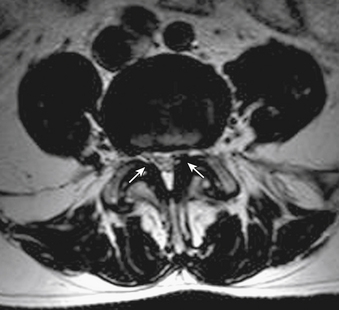
The ligamentum flavum takes the place of the joint capsule anteriorly and medially ligamentum flavum mri. Ligamentum flavum hematoma is difficult to diagnose preoperatively, even based on magnetic resonance imaging (mri).
Ligamentum Flavum Mri Anatomy: Ligamentum flavum hematoma is difficult to diagnose preoperatively, even based on magnetic resonance imaging (mri).

comment 0 Comments
more_vert